söndag 5 januari 2025

Mitokondrioitten virkistystä

 https://pubmed.ncbi.nlm.nih.gov/24606795/

Mitochondrial biogenesis: pharmacological approaches

Affiliations
Abstract

Organelle biogenesis is concomitant to organelle inheritance during cell division. It is necessary that organelles double their size and divide to give rise to two identical daughter cells. Mitochondrial biogenesis occurs by growth and division of pre-existing organelles and is temporally coordinated with cell cycle events [1]. However, mitochondrial biogenesis is not only produced in association with cell division. It can be produced in response to an oxidative stimulus, to an increase in the energy requirements of the cells, to exercise training, to electrical stimulation, to hormones, during development, in certain mitochondrial diseases, etc. [2]. Mitochondrial biogenesis is therefore defined as the process via which cells increase their individual mitochondrial mass [3]. Recent discoveries have raised attention to mitochondrial biogenesis as a potential target to treat diseases which up to date do not have an efficient cure. Mitochondria, as the major ROS producer and the major antioxidant producer exert a crucial role within the cell mediating processes such as apoptosis, detoxification, Ca2+ buffering, etc. This pivotal role makes mitochondria a potential target to treat a great variety of diseases. Mitochondrial biogenesis can be pharmacologically manipulated. This issue tries to cover a number of approaches to treat several diseases through triggering mitochondrial biogenesis. It contains recent discoveries in this novel field, focusing on advanced mitochondrial therapies to chronic and degenerative diseases, mitochondrial diseases, lifespan extension, mitohormesis, intracellular signaling, new pharmacological targets and natural therapies.

...

 Several pharmacological strategies to trigger these signaling cascades, according to these authors, are the use of bezafibrate to activate the PPAR-PGC-1α axis, the activation of AMPK by resveratrol and the use of Sirt1 agonists such as quercetin or resveratrol. Other strategies currently used include the addition of antioxidant supplements to the diet (dietary supplementation with antioxidants) such as L-carnitine, coenzyme Q10, MitoQ10 and other mitochondria-targeted antioxidants,N-acetylcysteine (NAC), vitamin C, vitamin E vitamin K1, vitamin B, sodium pyruvate or -lipoic acid As aforementioned, other diseases do not have exclusively a mitochondrial origin but they might have an important mitochondrial component both on their onset and on their development. This is the case of type 2 diabetes or neurodegenerative diseases. 

... In addition some of the current used strategies could be used to treat insulin resistance such as lifestyle interventions (caloric restriction and endurance exercise) and pharmacological interventions (thiazolidinediones and other PPAR agonists, resveratrol and other calorie restriction mimetics, AMPK activators, ERR activators). 

 ... Coenzyme Q10 and mitochondrial targeted antioxidants/peptides are reported to have the most remarkable effects in clinical trials. 

 They highlight a dual effect of PGC-1α expression on PD prognosis. Whereas a modest expression of this transcriptional co-activator results in positive effects, a moderate to substantial overexpession may have deleterious consequences. As strategies to induce PGC-1α activation, these authors remark the possibility to activate Sirt1 with resveratrol, to use PPAR agonists such as pioglitazone, rosiglitazone, fenofibrate and bezafibrate. Other strategies include the triggering of Nrf2/antioxidant response element (ARE) pathway by triterpenoids (derivatives of oleanolic acid) or by Bacopa monniera, the enhancement of ATP production by carnitine and alfa-lipoic acid.

 . A common hallmark of several neurodegenerative diseases (Huntington´s Disease, Alzheimer´s Disease and Parkinson´s Disease) is the impaired function or expression of PGC-1α, the master regulator of mitochondrial biogenesis.


Näyttöä koentsyymin Q10 hyödyistä silmälle viherkaihissa


doi: 10.4103/1673-5374.244781. Evidence on neuroprotective properties of coenzyme Q10 in the treatment of glaucoma
Affiliations

PMID: 30530997
PMCID: PMC6301180
DOI: 10.4103/1673-5374.244781 


The retina is one of the most metabolically active tissue in the body and requires an accurate regulation of the energy production. In this regard, mitochondria play a central role providing energy supply through oxidative phosphorylation. Adenosine triphosphate is generated by the electron transport chain complexes located within the cristae folds of the inner mitochondrial membrane. Moreover, mitochondria regulate a number of essential processes for neuronal functions including metabolic balance, intracellular calcium homeostasis, production of reactive oxygen species, and apoptotic signalling. Experimental studies have shown that RGCs death in glaucoma is an extremely complex process triggered by different molecular mechanisms (Nucci et al., 2018). Due to peculiar structural and energetic request, it is not surprising that RGCs are acutely vulnerable to mitochondrial dysfunction (Lee et al., 2011; Ito and Di Polo, 2017), which may increase their susceptibility to glaucoma-driven pathological factors (Figure 1). Main mechanisms of action of coenzyme Q10 (CoQ10).It has free radical scavenging property that prevents deoxyribonucleic acid (DNA) damage, has a fundamental role in cellular bioenergetics and a specific antiapoptotic activity by directly maintaining mitochondrial permeability transition pore (MPTP) in the closed conformation.

The specific loss of RGCs is a common feature of mitochondrial diseases (Lee et al., 2011). Indeed, inherited mitochondrial defects are associated with a number of optic neuropathies including Leber’s hereditary optic neuropathy and autosomal dominant optic atrophy, but also with more severe central nervous system (CNS) involvement in many other syndromic mitochondrial diseases, which are characterized by selective RGC death (Carelli et al., 2009; Lee et al., 2011; Ito and Di Polo, 2017).

The selective loss of neuronal populations and the cell damage pattern in glaucoma also resemble those of other neurodegenerative diseases (Nucci et al., 2016, 2018; Mancino et al., 2018a, b) and increasing evidence supports a causative role of the glutamate-induced excitotoxic mechanism in RGCs loss in in vitro and in vivo experimental conditions (Nucci et al., 2007). Several free radical scavengers and/or agents, that ameliorate mitochondrial function, have been candidate as treating agents to prevent cell death in various neurodegenerative conditions, such as Alzheimer’s disease and Parkinson, and glaucoma (Spindler et al., 2009; Ahmadinejad et al., 2017). CoQ10 is both a ubiquitous free radical scavenger and a recognised electron transporter in complexes I, II, and III of mitochondrial respiratory chain. CoQ10 is an important antioxidant and has a fundamental role in cellular bioenergetics. This led to consider glaucoma as a neurodegenerative disease and promoted clinical studies on new neuroprotective strategies not only targeted to IOP reduction (Nucci et al., 2016, 2018).

Interestingly, increasing evidence indicates that age-related mitochondrial defects play a central role in the pathogenesis of glaucoma (Nucci et al., 2007; Russo et al., 2008, 2009; Lee et al., 2014). Levels of CoQ10 in the human retina have been reported to declines with age (Qu et al., 2009). In this regard, it is well known the existence of a link between older age and the prevalence of glaucoma, thus suggesting a possible increased vulnerability of RGCs in glaucomatous neurodegeneration due to a lack of CoQ10 in older age (Bhagavan and Chopra, 2006; Lee et al., 2014).

This opens new opportunities of investigation for the development of novel neurotherapeutic agents for the treatment of glaucoma and other major retinal pathologies (Russo et al., 2008, 2009; Zhang et al., 2017).

CoQ10 poor aqueous solubility (Fato et al., 2010) and low bioavailability, partially due to the interactions with the multi-drug efflux pump P-glycoprotein expressed in both corneal epithelial cells and RGCs, have limited the development of topically active formulations of this molecule (Davis et al., 2017). To enhance the topical delivery and pharmacological effects of CoQ10, the co-administration with a P-glycoprotein inhibitor, such as D-α-Tocopherol polyethylene glycol 1000 succinate (TPGS), has been proposed (Davis et al., 2017). Interestingly, it has been successfully demonstrated both in vitro and in vivo mitochondrial-mediated neurotoxicity models that twice daily topical instillation of CoQ10/TPGS micelles was found to be significantly neuroprotective against RGCs loss than TPGS alone. The findings, in agreement with previous work, also suggest that the antioxidant activity of TPGS alone was insufficient to protect an immortalised neuronal cell line from insults generating mitochondrial oxidative stress, such as dimethyl sulfoxide and paraquat (Davis et al., 2017).

Topical ocular administration, in a model of high IOP–induced transient ischemia in rat, of CoQ10 0.1% + vitamin E (Vit E) 0.5% showed the ability to minimize DNA fragmentation and retinal cell apoptosis (Nucci et al., 2007) (Figure 1). This study confirmed, for the first time, that, during the reperfusion phase, the ischemic insult induces a significant increase of glutamate with consequent RGCs apoptosis. Thus, providing evidence of the usefulness of CoQ10 as a neuroprotective agent. In these conditions, administration of CoQ10 prevents glutamate increase, minimizing RGCs death in rats. It is plausible that the CoQ10 free radical scavenging mechanism may have a minor role in this process and that CoQ10 ability to restrains extracellular glutamate accumulation, may reduce the harmful effect of ischemia/reperfusion on mitochondrial energy metabolism and, accordingly, on the glutamate transporters function, preventing RGC apoptosis in the rats (Nucci et al., 2007). Excessive activation of glutamate receptors via the excitotoxic cascade leads to the MPTP formation and release of a proapoptotic factor, the cytochrome C, from the mitochondrial intermembrane space into the cytosol. Remarkably, CoQ10 inhibits this cascade by maintaining MPTP in the closed conformation, preventing apoptosis (Papucci et al., 2003).

The main concern about the topical administration was the concentration of CoQ10 at the retinal and vitreal level reached after the instillation of the eye drops. In this regard, it has been reported that when CoQ10 in association with Vit E eye drops are topically applied to the cornea, CoQ10 reaches the retina, substantially increasing local CoQ10 concentration and protecting retinal layers from apoptosis, in a mouse model of kainate-induced retinal damage. In addition, patients undergoing pars plana vitrectomy, who were administered CoQ10 in association with Vit E eye drops 1 hour before surgery, showed the presence of CoQ10 in the collected vitreous samples, thus confirming the ability of CoQ10 to reach the posterior ocular tissues (Fato et al., 2010; Lulli et al., 2012).

Oral administration of CoQ10 has also been reported to be neuroprotective in neurodegenerative diseases, as well as in cardiovascular diseases. CoQ10 supplementation has been reported to increase plasma CoQ10 concentrations, and the safety of high doses of orally-ingested CoQ10 over long periods has been well documented also in humans (Bhagavan and Chopra, 2006). Interestingly, it has been reported that CoQ10 is taken up by all tissues, including heart and brain mitochondria. This finding, together with growing evidence indicating that CoQ10 is neuroprotective in RGCs against IOP in vivo and in vitro, as well as against oxidative stress and excitotoxicity, suggests that CoQ10 could also be taken up by the retina and lead to a beneficial effect in glaucomatous retina (Lee et al., 2014). In a recent study on preglaucomatous DBA/2J and age-matched control DBA/2J-Gpnmb+ mice, diet supplementation with CoQ10 for 6 months was tested to assess the effects on glutamate excitotoxicity and oxidative stress-mediated RGC degeneration (Lee et al., 2014). Intriguingly, CoQ10 endorsed RGC survival, preserved the axons in the optic nerve head, and inhibited astroglial activation by reducing glial fibrillary acidic protein expression in the retina and optic nerve head of glaucomatous DBA/2J mice. Interestingly, CoQ10 significantly blocked the upregulation of N-methyl-D-aspartate receptor 1 and 2A, as well as of superoxide dismutase-2, heme oxygenase-1 protein expression in the retina of glaucomatous DBA/2J mice. Moreover, CoQ10 was able to prevent cell apoptosis by reducing Bax protein expression or by enhancing phosphorylated Bad protein expression. mtDNA content and mitochondrial transcription factor A/oxidative phosphorylation complex IV expression in the retina of glaucomatous DBA/2J mice were also preserved by CoQ10 supplementation. This suggest that CoQ10 may have a beneficial potential for ameliorating glutamate excitotoxicity and oxidative stress mediated glaucomatous neurodegeneration in the retina (Lee et al., 2014).

torsdag 2 januari 2025

Krystalliinigeenit ja proteiinit Silmän ihmeitä on läpinäkyvät, optista valoa taittavat kudokset.

 Aloitan katsoa näitä 2.1. 2025  GeneCards listasta.  (Etsin myös  löytöjä, miten voi  elämäntapaohjein ja ravinnon avulla  pitää huolta  silmänsä optisten kudosten pysymisestä  funktionaalisina.  Tässä on vasta  perustavaa pohjatietojen  etsintää 2.1. 2025)

https://www.genecards.org/Search/Keyword?queryString=Crystallins

(Mitä kystalliinit ovat? Ensimmäisestä krystalliinista sitaatti:Nisäkkäitten  silmän linssin krystalliinit on jaettu kolmeen perheeseen: alfakrystalliinit, beetakrystalliinit ja gammakrystalliinit. 

Alfakrystalliineja on kaksi geenituotetta , alfa-A ja alfa-B, asidinen ja baasinen. Ne muodostavat heterogeenisia aggrekaatteja (3:1), jotka toimivat ikäänkuin  kaitsijaproteiinit,  mutta niiden  tehtävänä  on pitää  eri proteiineja liukoisena. Alfa-A esiintyy silmän linssissä ja alfa-B voi esiintyä laajasti muissakin kudoksissa ja elimissä.  Silmän linssissä ei ole metabolista  turn over- tapahtumaa, joten  krystalliiniproteiineissa heijastuu ikämuutokset ja ikävaiheet. Krystalliinit ovat  olennaisia silmän linssin läpinäkyvyydelle.

NCBI Gene Summary for CRYAB Gene    (GeneCards)

  • Mammalian lens crystallins are divided into alpha, beta, and gamma families. Alpha crystallins are composed of two gene products: alpha-A and alpha-B, for acidic and basic, respectively. Alpha crystallins can be induced by heat shock and are members of the small heat shock protein (HSP20) family. They act as molecular chaperones although they do not renature proteins and release them in the fashion of a true chaperone; instead they hold them in large soluble aggregates. These heterogeneous aggregates consist of 30-40 subunits; the alpha-A and alpha-B subunits have a 3:1 ratio, respectively. Two additional functions of alpha crystallins are an autokinase activity and participation in the intracellular architecture. The encoded protein has been identified as a moonlighting protein based on its ability to perform mechanistically distinct functions. Alpha-A and alpha-B gene products are differentially expressed; alpha-A is preferentially restricted to the lens and alpha-B is expressed widely in many tissues and organs. Elevated expression of alpha-B crystallin occurs in many neurological diseases; a missense mutation cosegregated in a family with a desmin-related myopathy. Alternative splicing results in multiple transcript variants. [provided by RefSeq, Jan 2019]


    Muuta tietoa krystalliineista:

    https://pmc.ncbi.nlm.nih.gov/articles/PMC3554465/ 


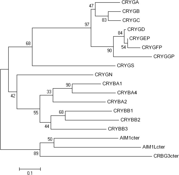
    Figure 3.

    Figure 3

    Phylogenetic tree of the humanβγ-crystallin gene superfamily. Sequences were extracted from the UCSC web browser. Translated ORFs were aligned and neighbor-joining trees were constructed as in Figure 2. AIM1, AIML, and CRYBG3 contain internal repeats corresponding to three β-crystallin-like genes in addition to regions not related to crystallin genes. For simplicity, the third, most highly conserved crystallin repeat from each gene was used for this alignment (designated AIM1cter, etc.).

    CRYB genes

    This multigene family has ancient origins in vertebrates, and indeed the six human genes have clear orthologs in fish []. β-crystallins are subdivided into acidic (A) and basic (B) subunits, encoded in CRYBA(12,4) and CRYBB(123) genes. Unlike the α-crystallins, four of the β-crystallin genes, arranged as two pairs, are close together on Chr 22 (Table 1, Figure 4).

    Table 1.

    Human lens crystallin genes

    Crystallin genes Chr
    α-crystallins
     
    CRYAA/αA
    21q22.3
    CRYAB/αB
    11q23.1
    β-crystallins
     
    CRYBA1/βA1, βA3
    17q11.2
    CRYBA2/βA2
    2q35
    CRYBA4/βA4
    22q12.1
    CRYBB1/βB1
    22q12.1
    CRYBB2/βB2
    22q11.3
    CRYBB2 P1
    22q11.3
    CRYBB3/βB3
    22q11.23
    γ-crystallins
     
    CRYGAA
    2q34
    CRYGBB
    2q34
    CRYGC/γC
    2q33.3
    CRYGD/γD
    2q33.3
    CRYGEPE
    2q33.3
    CRYGFPF
    2q34
    CRYGS/γS
    3q27.3
    CRYGNN
    7q36.1
    CRYGGP
    2p16.3
    ‘Enyzme-crystallin’
     
    BHMT/ψ 5q14.1

    Gene/protein names and chromosome locations are shown. Genes/proteins in bold type are expressed at relatively high levels similar to orthologs in other mammals. Those in normal type seem to have reduced or no expression in human lens. Some are designated pseudogenes. The proposed crystallin designation for betaine-homocysteine methyltransferase (BHMT), ψ, does not imply pseudogene status.

Onko olemassa terapiaa joka estää kaihia muodostumasta oksidatiivisessa stressissä?


Review
doi: 10.3390/antiox13101249.Oxidative Stress in Cataract Formation: Is There a Treatment Approach on the Horizon?
Affiliations
Abstract

Cataracts, a leading cause of blindness worldwide, are closely linked to oxidative stress-induced damage to lens epithelial cells (LECs). Key factors contributing to cataract formation include aging, arterial hypertension, and diabetes mellitus. Given the high global prevalence of cataracts, the burden of cataract-related visual impairment is substantial, highlighting the need for pharmacological strategies to supplement surgical interventions. Understanding the molecular pathways involved in oxidative stress during cataract development may offer valuable insights for designing novel therapeutic approaches. This review explores the role of oxidative stress in cataract formation, focusing on critical mechanisms, such as mitochondrial dysfunction, endoplasmic reticulum stress, loss of gap junctions, and various cell death pathways in LECs. Additionally, we discuss emerging therapeutic strategies and potential targeting options, including antioxidant-based treatments.

Keywords: aging; antioxidants; cataract; diabetes; hypertension; novel treatment approaches; oxidative stress.


Figure 2
Illustration of the central pathogenetic pathways during cataracto-genesis associated with aging, systemic hypertension or diabetes. ATP: adenosintriphosphat; Ca2+: calcium; ER: endoplasmic reticulum; GSH: glutathione; K+: potassium; mtDNA: mitochondrial DNA; Na+: sodium; NOX: nicotinamide adenine dinucleotide phosphate oxidase; ROS: reactive oxygen species; UPR: unfolded protein response. Upward arrows indicate upregulation or increased activity or increased concentration, whereas downward arrows indicate downregulation or decreased activity or decreased concentration.


Katse katseeseen, silmäys silmään... Miten estää kaihia?

 Ferulihappo mainitaan tällä ajattelun linjalla. Onkohan asia *koeputkitasolla vai koe-eläintasolla?? Joku mainitsee ferulihapon "antioksidanttien antioksidanttina".

https://onlinelibrary.wiley.com/doi/epdf/10.1002/jbt.70031

RESEARCH ARTICLE
Ferulic Acid Protects Human Lens Epithelial Cells Against UVA-Induced Oxidative Damage by Downregulating the DNA Demethylation of the Keap1 Promoter
First published: 29 October 2024
Xinru Ling and Lei Zhu are the co-first authors of this study.
Abstrakti sitaattina: 

ABSTRACT

Ultraviolet (UV) radiation-triggered production of reactive oxygen species (ROS) is a primary contributor to apoptosis in human lens epithelial cells (HLECs), which can ultimately result in cataract formation. The nuclear factor erythroid-2-related factor 2 (Nrf2)-Kelch ECH associating protein 1 (Keap1) pathway, a fundamental oxidative stress regulation mechanism, plays a crucial role in the development of cataracts.

 Ferulic acid (FA), recognized for its potent antioxidant properties can activate the Nrf2 signaling pathway to mitigate oxidative damage and cell apoptosis. In this study, we have demonstrated the protective effects of FA in reducing UVA-induced oxidative damage and apoptosis in HLECs through the modulation of the Keap1/Nrf2 pathway, as evidenced by both cellular and animal experiments. HLECs and Lens* were exposed to 10 J/cm2 UVA radiation with or without prior treatment with FA. We found that UVA radiation increased oxidative damage and cell apoptosis in HLECs, ultimately leading to opacification of rat lenses, while FA was able to attenuate both oxidative damage and cell apoptosis in HLECs and reduce the degree of lens opacification. FA upregulated the expression of antioxidant response factors of the Keap1/Nrf2 pathway and downregulated the expression of apoptosis-related genes in HLECs, as demonstrated by Western blot and RT-qPCR analyses. We also found that UVA radiation increased the degree of demethylation of the Keap1 promoter in HLECs, whereas FA reduced the level of Keap1 promoter demethylation as determined by DNA sequencing. Additionally, UVA upregulated the expression of DNA active demethylase of the Keap1 promoter in HLECs, Dnmt1, Dnmt3a, and Dnmt3b, as shown by immunofluorescence, Western blot, and RT-qPCR, however, FA attenuated the activity of the passive demethylase TET1 in addition to the active demethylases. These results demonstrated that UVA radiation can cause oxidative damage, cell apoptosis, and rat lens opacification by increasing the demethylation of the Keap1 promoter in lens epithelial cells. Conversely, FA was shown to reduce oxidative damage, inhibit cell apoptosis, and decrease rat lens opacification by increasing the methylation of the Keap1 promoter. These findings suggest that FA could be therapeutically beneficial in preventing and mitigating cataracts induced by UVA radiation.

FERULIHAPPO

Feruliinihapon toimittajat-VIHREÄ KEVÄT®

Green Spring on ammattimainen luonnollisen ferulihapon valmistaja. Tuottamamme luonnollisen feruliinihapon pitoisuus voi olla 98,7 % uutettuna kasviperäisistä riisileseistä, ja jäännösstandardi on paljon alhaisempi kuin eurooppalainen standardi, jota voidaan käyttää laajasti elintarvikkeissa, lääkkeissä, terveydenhuoltotuotteissa ja kosmetiikassa. Voimme tarjota sinulle korkealaatuisia tuotteita parhaaseen hintaan ammattimaisena luonnollisena ferulihapon toimittajana,Vihreä keväton paras valintasi.

Feruliinihapon kemiallinen nimi on 4-hydroksi-3-metoksikanelihappo, joka on yksi kanelihapon johdannaisista (tunnetaan myös nimellä kanelihappo, 3-fenyyli-2-akryylihappo, molekyylirakenne). Ferulihappoa löytyi alun perin kasveista. Se löytyy kasvin siemenistä ja lehdistä, ja se on fenolihappo, jota esiintyy laajalti kasveissa. Se yhdistyy soluseinän polysakkaridien ja proteiinien kanssa muodostaen soluseinän luuston.

Sillä on korkea pitoisuus kiinalaisissa lääkeaineissa, kuten Asafoetida, Angelica, Chuanxiong, Cimicifuga, Jujube-siemen jne. Se on yksi näiden kiinalaisten lääkkeiden tehokkaista ainesosista ja sitä on käytetty yhtenä kiinalaisten patenttilääkkeiden laatuindikaattoreista. Elintarvikkeiden raaka-aineista myös kahvi, akanat, pandan-pavut, vehnäleseet ja riisileseet sisältävät enemmän ferulihappoa. Farmakologisten vaikutusten tutkimukset ovat viime vuosina löytäneet feruliinihapon ja sen johdannaisten monia farmakologisia vaikutuksia ja biologisia aktiivisuuksia, ja myrkyllisyys on alhainen, joten sillä on laaja käyttöalue lääketieteessä, terveydenhuollon tuotteissa, kosmeettisissa raaka-aineissa ja elintarvikelisäaineissa. .


lördag 23 november 2024

Suoliston mikrobiomin osuudesta terveydessä Ihmisen toinen genomi. Probiootit, prebiootit...

 NAFLD Review    , Oirealue "Steatorrhea", rasvaripuli, kelluvat ulosteet ja  suoliston  toiminnanhäiriö

Ilmeisesti vaivaa  maailmankansoja, koska sanotaan 25% aikuisista olevan näitä  oireita. Terapiaa  etsitäänkin   dieetin, funktionaalisen ravinnon, elämäntapojen ja liikunnan   alueilta enemmön kuin  ihmisen omien geenien. Suoliston microbiomia sanotaan ihmisen toiseksi geenistöksi, niin paljon sieltä löytyy geneettisen  rakenteen omaavaa   elävää yksilöllistä eliötä.  Onhan ihmisen mitokondrioillakin, jotka ovat joskus bakteereista  jo tulleet ihmiseen, jotain sanottavaa  ihmisessä. Mitokondriat nykyisellään  ovatsopeutuneet organelleja  hyödyttämässä  ihmistä  geneettisenä lisälaitteena   Ne periytyy äidin puolelta koska munasoluissa niitä mahtuu olemaan  seuraavalle  sukupolvelle  alkulahjana.   Jotkut  mikroeliöt vieläkin  asettavat  ihmisen genomiini jotain uutta- kuka tietä mitä ihmeitä  valtava mikroeliö maailma  asettaa ihmissukupolviin. Joka tapauksessa  yksi provirus-ilmiö on tarkasti selvittetty ( HIV viruksen  tekemä pro-virus jää ihmisen DNA:han. Miten sitten meioosissa?

  Mutta  nyt on kyse  auoliston sisällä vielä aivan itsenäisenä elävistä  bakteerilajeista ja niiden keskinäisestä elämäntaistelusta. Toiset  ovat  hyödyllisempiä ihmisen itsensä suhteen kuin toiset lajit ja ihminen voi elintavoilla, ravinnolla , liikunnalla vaikuttaa  omaan  suolistomikrobiomiinsa jonkin verran.   Tämä  alue on  laajan tieteellisen tutkimuksen kohdetta yhä edelleen ja suolisto-ongelmat puolestaan ovat  enenemään päin  globaalisti.   Ihmisen täytyy ottaa tiedon lisääminen yhdeksi aseekseen terveytensä puolesta.  Mikrobeillä on luonnollinen evoluutionsa ja eskalaationsa.

Nonalkoholinen rasvamaksatauti (steatorrhean  taustalla)

https://pubmed.ncbi.nlm.nih.gov/?term=nafld+review&sort=date

 pro-biootti, pre-biootti, synbiootti, post-biootti,

ovat tämän alueen sanastoja.   

https://www.wjgnet.com/1007-9327/full/v30/i43/4682.htm

The gut-liver axis plays a crucial role in the development and progression of metabolic dysfunction-associated steatotic liver disease (MASLD). Key metabolites, including lipopolysaccharides, short-chain fatty acids (SCFAs), bile acids, and beneficial gut bacteria such as Bifidobacterium and Lactobacillus, are pivotal in this process. Glucagon-like peptide-1 receptor agonists (GLP-1 RAs) show promise in managing MASLD by promoting weight loss, enhancing insulin secretion, and improving liver health. They restore gut-liver axis functionality, and their effects are amplified through dietary modifications and gut microbiome-targeted therapies. Emerging research highlights the interplay between GLP-1 RAs and gut microbiota, indicating that the gut microbiome significantly influences therapeutic outcomes. Metabolites produced by gut bacteria, can stimulate glucagon-like peptide-1 (GLP-1) secretion, further improving metabolic health. Integrating dietary interventions with GLP-1 RA treatment may enhance liver health by modulating the gut microbiota-SCFAs-GLP-1 pathway. Future research is needed to understand personalized effects, with prebiotics and probiotics offering treatment avenues for MASLD. Key Words: Metabolic dysfunction-associated steatotic liver disease; Glucagon-like peptide-1 receptor agonists; Gut microbiome; Gut-liver axis; Diet intervention

Guney-Coskun M, Basaranoglu M. Interplay of gut microbiota, glucagon-like peptide receptor agonists, and nutrition: New frontiers in metabolic dysfunction-associated steatotic liver disease therapy. World J Gastroenterol 2024; 30(43): 4682-4688 [PMID: 39575401 DOI: 10.3748/wjg.v30.i43.4682]

tisdag 25 april 2023

Luun uudismuodostukseen vaikuttavista öljyistä


. 2020 Aug 19;11(8):7048-7060.
doi: 10.1039/d0fo00884b.

The opposite effects of Antarctic krill oil and arachidonic acid-rich oil on bone resorption in ovariectomized mice

Affiliations
  • PMID: 32813003
  • DOI: 10.1039/d0fo00884b 
  •   Osteoporosis, a chronic disease that affects over 200 million people worldwide, presents a substantial medical and socioeconomic burden on the modern society. However, long-term intake of diets supplemented with different polyunsaturated fatty acids (PUFAs) can affect bone metabolism; thus, this study investigated the comparative effects of Antarctic krill oil (AKO, containing n-3 PUFAs) and arachidonic acid-rich oil (AAO, containing n-6 PUFAs) on bone resorption in a mice model of postmenopausal osteoporosis. Mice were orally administered with AKO (200 mg kg-1) or AAO (220 mg kg-1) once daily for 30 days, ovariectomized, followed by the continued administration of the respective samples for 90 days. Biomechanical and histomorphometric analyses revealed that AKO increased the bone mineral density (BMD) to enhance the biomechanical properties by increasing the mineral apposition rate and repairing the microstructure of the trabecular bone, whereas AAO had the opposite effect. The fatty acid analysis of the vertebra showed that AKO increased the n-3 PUFA (especially for DHA) content, thereby decreasing the ratio of n-6/n-3 PUFAs, which was negatively correlated with the BMD. However, AAO had the opposite effect due to high amounts of arachidonic acid. To explore the underlying mechanism responsible for these observations, we compared the classical bone resorption OPG/RANKL/NF-κB pathway mediated by PGE2/EP4. The ratio of n-6/n-3 PUFAs in the bone affected the production of PGE2, a factor regulating the OPG/RANKL pathway, thereby regulating osteoclastogenesis by stimulating the NF-κB pathway. The results of ELISA, qRT-PCR, and western blot demonstrated that AKO reduced the secretion of PGE2 and the expression of EP4, upregulating the ratio of OPG/RANKL in the bone, thereby decreasing TRAF6 expression to inhibit the activation of the NF-κB signaling pathway, and finally inhibiting the expression of nuclear transcription factors (c-fos and NFATc1) to prevent excessive osteoclastogenesis (TRACP, MMP-9, and Cath-K). Arachidonic acid is a precursor of PGE2 synthesis. AAO showed the opposite trend through the same pathway. Thus, AKO could significantly improve osteoporosis via the OPG/RANKL/NF-κB pathway mediated by PGE2/EP4 to inhibit osteoclastogenesis, whereas AAO aggravated osteoporosis via the same pathway. This is the first study to systematically compare the effects and mechanism of AKO and AAO in regulating bone resorption in osteoporotic mice to support recommendations on fatty acid types in dietary oils for an osteoporotic population.